Hip arthroscopy
The hip joint is deep inside the body and is difficult to access and investigate. Often people have hip pain which cannot be diagnosed with traditional tests and may be left untreated until recognisable and irreversible damage has occurred.
Until recently, most procedures on the hip joint involved open surgical dislocation of the joint with a significant hospital stay and rehabilitation. The advances in minimally invasive arthroscopic techniques has lead to early treatment of young active patients and those with sporting careers ahead of them.
Treating some hip problems early using arthroscopy can help delay the need for joint replacement which would be required sooner if left untreated.
During the procedure minimally invasive tools and technique are used to examine the hip and to correct problems through small incisions rather than through more invasive surgery.
The procedure itself involves a small number of incisions being made. A miniature high-definition camera is then inserted into one portal and other portals are used as required for instruments.
Download pdf patient information guide on hip arthroscopy
Advances in instrument technology and techniques have improved access to the joint and enabled greater scope of treatment.
Femoroacetabular Impingement – FAI
The hip joint is a type of ball and socket joint which is made of the the femoral head (the top of the thigh bone which is shaped like a ball) and the acetabulum (part of the pelvis which forms a socket for the ball to sit in).

Femoroacetabular Impingement (also shorted to FAI) occurs when either the ball or the socket are abnormally formed.
This results in the ball and socket rubbing abnormally against each other. This causes the hip joint to wear out, causing damage to structures that are important to the proper function of the hip such as:
- cartilage damage
- labral tears
- ligamentum teres tears
- and eventually osteoarthritis of the hip.
FAI is a common condition, affecting 25% of people and is one of the most common causes of hip arthritis.
There are 2 types of FAI.
-
CAM impingement. The bone abnormality is located on the ball.
-
PINCER impingement. The bone abnormality is located on the socket.


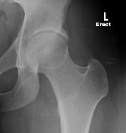
Left– Normal hip XR in frontal view:
Right– Pincer Impingement, XR in frontal view:
Below – Combined Pincer & Cam impingement, XR in Dunn (rotated)view

Appearance of normal XR in frontal view

Left– Normal hip XR in frontal view
Right– Hip appears to look normal on CT 3D reconstruction in the frontal view
Below – Same hip BUT Cam lesion clearly visible on CT 3D reconstruction in the Dunn (rotated) view

Labral tears

The labrum is a special type of cartilage which forms a rim around the hip socket (acetabulum). It helps keep the joint stable and allows the hip joint to function properly. It acts similar to a wet suction cup when it is pulled or slid along a window pane. A key property is that when it is functioning properly it prevents synovial fluid leaking out of the joint.
Synovial fluid has several functions and is very important to the the long term health of a hip joint and prevention of early arthritis. By acting as a boundary lubricant it decreases abrasive contact between articular cartilage of the femoral head and acetabulum.
Synovial fluid is also a critical nutirent that keeps articular cartilage alive. Articular cartilage has no arteries, veins or nerves. As it is irreversibly damaged no pain is felt until either the bone underneath is exposed or other tissue that is supplied with nerves in the joint is inflammed.
The labrum however has a nerve supply and when torn it causes hip and deep groin pain and unfortunately may not heal naturally by itself. It can cause a catching sensation as the labrum is “caught” within the hip joint “jamming” into articular cartilage damaging it until released. If the labral tear is large it may remain “jammed” giving a locked sensation of the hip for several days until it is released. In this situation it is important to see your orthopaedic surgeon to avoid significant irrevesible cartilage damage.

Blue arrow – inflammed hip capsule ( mild synovitis )
Black arrow – labrum
Red arrow – labral tear with separation from acetabular rim cartilage ( labrochondral juction also torn)
Red star – femoral head

Black arrow -gentle pressure on torn labrum
Red arrow – bubble of acetabular cartilage as it separates from bone

Black arrow – labrum repaired in anatomical position and under pressure
Blue arrow – Labrochonrdal junction anatomically recreated and stable.
Red arrow – Cartilage reduced and stabilised with labral repair. Microfracture to acetabular bone ensures cartilage rebonds. In situations where cartilage flaps occur and not stabilised with labral repair fibrin glue can be used.
It is important to realise the differences between a hip labral tear from a knee meniscal tear. The labrum is attached to the hip cartilage while the meniscus is not attached to knee cartilage. So if the labrum is torn there has been irreversible damageto hip cartilage which usually does not occur with meniscal tears.
A labral tear may heal with conservative management in 6-8 weeks. The cartilage may heal with scar tissue formation. The scar tissue is fibrocartilage and not articular hyaline cartilage.


Ligamentum Teres Tears
The ligamentum teres, is a triangular, coiled and flattened ligament that connects the ball (femoral head) to the socket (acetabulum) of the hip joint. It is similar in function to the anterior and posterior cruciate ligaments to the knee.


The ligament teres is made tense when the thigh is semiflexed and the limb then abducted or rotated outward. It is the last soft tissue restraint important for stability preventing your hip from dislocating. As a result it is a commonly injured in those who regularly force their hips into uncommon positions.
To achieve some dance and yoga positions (e.g.splits or lotus position) the hip has to to subluxate (partially dislocate). Subluxation can only occur if the ligament can stretch sufficiently. When the ligamentum teres tears patients often describe a popping sensation followed by pain that can last 2 to 6 weeks. The hip pain may resolve and healing occurs with scar tissue. The healed but scarred ligament teres is now less elastic and risk of retear is greater. Especially for those who regularly stretch the ligament and develop a cycle of pain and recovery with retears.
Diagnosis of a ligamentum teres tear (above) is difficult . This is based on clinical history, signs and symptoms. The ligamentum teres is important for propioception and innervated with a large nerve supply. Those with a tear often describe a sensation that their hip is “dislocating” or “coming apart”.
Patients may not initially have hip pain until advanced irreversible damage has occurred. The complex nerve innervation of the ligamentum teres results in referred pain , particularly to lower back, anterior pelvic, buttock, thigh and leg. This makes it difficult for patients to describe.
Patients often have failed multiple treatments over several years until diagnosed. Pain can mimic sciatica (spinal nerve impingement), gynaecological symptoms (eg endometriosis) and abdominal symptoms (eg appendicitis). Some patients have had failed surgery such as appendectomy but this is becoming rare as more is learnt about presentation.
Imaging is poor for diagnosing ligamentum tears. MRI will miss 50% of ligamentum teres tears. This is primarily becuase the ligament is not stretched when you are lying flat inside the MRI . To be in a position where the ligamentum teres is stretched you would not fit inside the MRI tube.
However we are fortunate that we can inject the hip joint fluroscopically with local anaesthetic and steroid. The local anaesthetic will relieve all pain both direct and referred that is coming only from within the hip joint. This does not imply that the LT is torn but informs us if the problem is within the hip joint or that we need to keep investigating for a problem elsewhere (e.g. tendon, bursa, muscle, nerve or spine).
Patients are asked to keep a pain diary before and after the injection to differentiate the for short term (local anaesthetic) and long term (steroid) changes. It is crucial that pain must be significant, around 7 out of 10, before the injection as the local anaesthetic can not numb something that is not painful. Download pain diary pdf.
Ligamentous laxity & Ligamentum teres tears
Ligaments are elastic and are naturally able to stretch. The ability of a person to “stretch” or describing themselves as “naturally flexible” relies on a combination of the mechanics of your joints and the elasticity of your ligaments. However, flexibility most commonly comes from the elasticity of your ligaments.
Most people have to “train” their ligaments to improve their elasticity with regular stretching and exercise. Others have “natural flexibility” which is a spectrum from being somone who always was “flexible” irregardless of exercise to those who could always achieve hypermobilie positions easily (e.g. splits). It is often these people who have laxity.
Some are occasionally sufficiently lax they can partially dislocate (subluxate) and in some cases even disolocate their joints without difficulty. Unfortunately ligaments with increased elasticity are more likely to lengthen and tear with repeated subluxation. Just as occurs with repeated stretching of an elastic band.
Repeated tearing and lengthening of an abnormally lax ligament further increases laxity and instability of the joint compounding the problem.
Partial dislocation of the femoral head causes point loading onto the labrum and hip cartilage. Note in the video above from the start the hip is already partially dislocated anteriorly without forced rotation and the origin of the ligamentum teres on the femoral head is touching the labrum which is not normal. The labrum is the lontitudinal band of white tissue in the centre of the image, the femoral head is on the right and the anterior acetabular cartilage is on the right. With increased external rotation of the hip the the femoral head can be seen to partially dislocate further and the abnormal LT can be seen to be significantly lengthened and unable to prevent subluxation. Repeated subluxation such as this can result in early irreversible damage and osteoarthritis.
In the early stages before significant ligamentum teres, labral or cartilage damage occurs all imaging of a ligamentously lax joint is normal. As imaging is not dynamic (e.g. MRI) the hip is not forced to subluxate and so all appears normal. Ligamentous laxity also known as joint hypermobility has a series of simple screaning tests (Wynne Davies Ligamentous Laxity tests JBJS 1970). However this will often miss those with mild to moderate laxity.
Wynne Davies criteria
If 3 of the 5 pairs of joints examined in any one individual demostrate this degree of laxity it is taken as positive1. Thumb touching forearm on flexing wrist
2. Fingers parallel to forearm with wrist extension
3. Elbows extend past 180°
4. Knees extend past 180°
5. Foot dorsiflex past 45°
Diagnosing and managing ligamentous laxity is crucial to preventing early irreversible damage. This is done in conjunction with your physiotherapist by strengthen muscles, protecting range of motion and avoiding position which cause partial dislocation. It is difficult for some to stop specific sport or activities and they may continue to subluxate their hip with resulting significant risk of injury.
Chronic ligamentum teres tears
Chronic ligamentum teres tears that do not heal can cause damage to hip cartilage (arthritis) in the non weightbearing areas of the hip joint (medial wear). This is caused by chronic grating of the torn ligament in between the cartilage lining the femoral head and acetabular socket. Medial wear will only present on plain XRs when advanced arthitis is present and not salvageable by arthroscopy.


Chronic inflammed and haemorrhagic ligamentum teres tear with significant inner (medial) femoral head cartilage wear (advanced arthritis).
Note
Left image the large loose cartilage body found floating within the hip joint and significant cartilage damage.
Right image of the same femoral head demonstrating where the loose body came from and a chronic cartilage flap soon to tear free and become a second large loose body within the hip joint. Each loose body within the hip joint propogates and accelerates damage leading to arthritis.
The above patient presented with 4 years of undiagnosed lower back and anterior pelvic pain and recent rapid progression investigations with normal CT and MRI hip and spine and gynaecological review and work up. Fluroscopically guided hip joint injection was diagnostic resolving referred pain indicating that the problem was within the hip joint and not elsewhere. The ligament teres tear and loose bodies were not diagnosed until arthroscopy was performed.
The ligament teres as discussed earlier is an important stabiliser in extreme ranges of motion. If the ligament is torn and does not heal the chronic tear can result in instability and the hip will subluxate if the movement/exercise which tore it is continued. If this occurs for a long period (greater than 3 months) the ligamentum teres debris will rub on acetabular cartilage wearing it away resulting in arthritis and pain.

Ligament teres intrasubstance tear apparently insignificant when hip in neutral position.

Same hip placed in Internal rotation demonstrating the tear is greater than initally seen. There is cartilage damage to the femoral head where the tear is constantly rubbing.

Same hip after radiofrequency ablation of the torn fragments visible. Approximately 50% of ligamentum teres remaining.
Dynamic view of same hip as rotation occurs from internal to external rotation. Note the trough of cartilage damage in the acetabulum where the ligamentum teres tear had been abraiding if the hip continued to externally rotate and subluxate. In this demonstration the hip was not forced into complete subluxation to prevent remaining ligamentum teres causing further cartilage injury.
It is important to discuss your symptoms with physiotherapist or sports physician. As a general rule most soft tissue injuries should have helaed within 6 weeks and completely recovered by 12 with appropriate conservative management. If symptoms continue it is likely a chronic injury has developed that cannot heal itself.
Chondroplasty & Loose Bodies
Acute cartilage delamination and flap formation
Occasionally hip cartilage can tear traumatically and presents differently than a similar injury in the knee . Hip joint cartilage delaminates (ie “separates like orange peel”) from the bone more easily than the knee.
Delamination results in the formation of a mobile cartilage flap. A cartilage flap is mechanically self propagating and may rapidly increase in size with associated pain despite normal/reduced activity. This is different to slowly progessive arthritis where the cartilage is gradually worn away over years.
A loose body within the joint can present similarly to acute cartilage flap formation and is managed by arthroscopic removal. These lumps of cartilage, or bone and cartilage, can gradually grow larger within the joint. These can jam and cause locking symptoms and severe pain, and may cause joint inflammation (synovitis). Over time, they may cause damage to the joint cartilage and lead to the development of osteoarthritis.
When a flap is formed arthroscopically it can be trimmed to stable edges (Chondroplasty). This leaves an area of full thickness cartilage loss and exposed bone in the hip joint. The exposed bone is microfractured allowing mesenchymal stem cells from the pelvic marrow to be released and form a blood clot. These stem cells in turn increase the chance of fibrocartilage scar formation covering the exposed bone.
Fibrocartilage scar takes several months to mature and adhere. It is not as strong as the previous hyaline cartilage which was lost. As a result it is advised to avoid high impact exercise as this could dislodge the scar tissue with return of pain and progressive damage.
If the cartilage flap formed recently, is in good condition and not degenerate it may be possible to bond it back to the acetabular bone with a combination of fibrin glue and microfracture. If this fails a repeat arthroscopy is required to trim the remaining unstable cartilage. Despite the risk of possible repeat surgery this is my prefered method as hyaline cartilage is far superior than fibrocartilage scar tissue. If repeat surgery is needed usually the majority of flap has united with the bone leaving a smaller area exposed.
Below arthroscopic images of a patient with sudden onset of hip pain and loss of function from minimal trauma. Pain was progressive over 3 months and unable to work. MRI report of moderate arthritis and little else of note. Arthroscopy demonstrated the moderate arthirits initially (Left). But a massive mobile cartilage flap covering 50% of weight bearing area of hip joint became visible when probed which was causing the symptoms (Right).


Below after debridement of the flap and microfracture is performed on the exposed bone (Left). Bleeding and release of bone marrow and stem cells can be seen from areas of microfracture (Right).


Note: Because of the relatively normal MRI but significant pain a fluoroscopically guided hip joint injection was indicated. This injection was diagnostic relieving his pain, albeit briefly. This confirmed that his symptoms were from inside his hip joint, not elsewhere and that a hip arthroscopy was appropriate.
Loose bodies
A loose body within the joint can present similarly to acute cartilage flap formation and is managed by arthroscopic removal. These lumps of cartilage, or bone with cartilage attached, can gradually grow larger within the joint. These can jam and cause locking (mechanical) symptoms and severe pain, and may cause joint inflammation (synovitis). Over time, they may cause damage to the joint cartilage and lead to the development of osteoarthritis.
An acute traumatic injury to the hip, such as hip dislocation, can create multiple loose bodies of cartilage that still have bone attached that float around the joint. This should be suspected if a hip continues to have mechanical symptoms following dislocation despite a “normal” report of a CT or MRI as can be easily not visualised
Preventing re-injury
When there has previously been painful areas of full thickness cartiIage loss and exposed bone I would not recommend returning to high impact exercise, particularly running on hard surfaces, until symptom free for up to 12 to 18 months following arthroscopy.
To lower your chances of re-injury I recommend addressing three things before return to exercise :
Footwear
Poor footwear could be old, worn-out shoes that need replacing (running shoes need replacing at about the 750km mark) or new shoes may have the incorrect support. Invest in supportive shoes designed for your unique foot structure to optimise shock absorption. A podiatrist can help you with the choice.
Core strength
Core strength is key to any physical activity. A strong core gives stability to your torso and acts as a buffer. Working with a physiotherapist or personal trainer to build up your core strength is crucial and should be strong particularly when returning after recovering from surgery.
Overtraining
As a general rule the majority of pain and injuries are the result of simple overtraining. As a guide increase your training distance by a maximum of 10% each week provided you continue to remain asymptomatic.
Osteoarthritis & Hip Arthroscopy
Those with arthritis and associated mild discomfort and symptoms over many years can develop sudden onset progressive pain for no apparent reason. In the situation of advanced arthritis a salvage arthroscopy is unlikely to be of more than short term benefit other than to document the extent of cartilage loss that may not have been apparent on an XR and MRI.
As a general rule if there is less than 2mm of joint space between the ball (femoral head) and socket (acetabulum) of your hip joint arthroscopy is unlikely to provide any significant benefits in the longer term.


The Progressive cartilage degeneration which occurs over many years, although fundamental to the cause of osteoarthritis, is not the site of origin of pain. Peripheral nerves generally follow the path of blood vessels, and cartilage contains no nerves or blood vessels. Pain may be from the joint capsule , synovium (lining of capsule which produces fluid) , ligamentum teres and labral degenerative tears (as discussed above but chronic) or bone as well as in the muscles and soft tissues, which surround and move the joint.
Trochanteric Bursitis
Greater Trochanteric Pain Syndrome is the name given to pain felt about the outer (lateral) side of the hip. It has previously had many other names, including trochanteric bursitis (inflamed painful bursa), and gluteal tendinopathy (painful typically Gluteus Medius tendon microscopic partial tears).
It most commonly effects women over 40 years of age, but can also occur in younger athletes. The associated pain can be very severe. It will often be worse at night, but also when walking on hills and steps. There will generally be marked local tenderness when pressure is applied to the side of the hip.
GTPS is treated initally once diagnosed with physiotherapy, and may require one or several cortisone injections. A single injection of Leukocyte rich PRP shows promise as more effective treatment than repeated cortisone injestions. If the Gluteal tendons are found to torn and the cause of ongoing pain, then surgical repair is likely to be required.

When conservative management has been optimised and failed it is known as recalcitrant GTPS. We have published a paper on the results of 49 of our patients following arthroscopic surgery which available to down load – The outcome of endoscopy for recalcitrant greater trochanteric pain syndrome. Drummond J, Fary C, Tran P. Arch Orthop Trauma Surg. 2016 Nov;136(11):1547-1554.
Psoas Tendinitis/Bursitis
The tendon of the Psoas muscle sits in the front of the hip joint. It is most often injured by overuse or overload, but these injuries will most often respond well to physiotherapy treatment.
The tendon can be injured by rubbing against a rough surface such as a bone spur (osteophyte), or the edge of a total hip replacement socket. Such damage will cause pain in the front of the hip but this can be difficult to differentiate from pain generated by to surrounding soft tissue and bony structures nearby. Psoas injury is associated with marked weakness of hip flexion. This may lead to great difficulty climbing stairs, for example.
The diagnosis of Psoas tendinitis is confirmed with an ultrasound examination, plus injection of local anaesthetic and cortisone around the tendon. The injection should give temporary improvement in symptoms. Permanent resolution of symptoms can generally be obtained by arthroscopic tendon lengthening. This surgery may sometimes be associated with some weakness hip flexion, but this nearly always recovers.
Snapping Hip Syndromes
The sensation or audible sound of snapping around the hip may be felt during hip movements. There are two types of snapping. “Internal snapping” usually an audible snap, or clunk, when the hip is extended from a flexed position. This is caused by the Psoas (a major hip flexor, in the front of the hip) tendon catching while sliding over the hip joint and then suddenly giving. Typically patients will describe it as their “party trick” until it becomes painful. “External snapping” is often not audible, but is felt by the patient on the side of the hip.
Both forms of snapping may be treated with cortisone injections to try to settle inflammation and swelling, and physiotherapy to try to release tightness in the muscle and tendon. If these treatments are unsuccessful, and the symptoms warrant, then arthroscopic surgery may be performed to lengthen the tendons and prevent further catching. This surgery successfully relieves snapping in the great majority of cases.
For more information on hip arthroscopy :
- Surgery and the conditions treated : SOS Clinic website
- Evidence based medicine : Up-to-date resource on hip arthroscopy research
- International Society for Hip Arthroscopy

You must be logged in to post a comment.